

Julio A. Aguirre-Ghiso, Ph.D.
- Professor, Department of Cell Biology
- Professor, Department of Oncology (Medical Oncology)
- Professor, Department of Medicine (Oncology & Hematology)
- Rose C. Falkenstein Chair in Cancer Research
- Co-Leader, Montefiore Einstein Comprehensive Cancer Center, Tumor Microenvironment & Metastasis Program
- Director, Cancer Dormancy Institute
Area of research
- Metastasis biology , Cancer cell dormancy , Signal transduction , Extracellular matrix , Innate immune cell function , tumor and normal organ microenvironment
Phone
Location
- Albert Einstein College of Medicine Michael F. Price Center 1301 Morris Park Avenue 220 Bronx, NY 10461
Research Profiles


Professional Interests
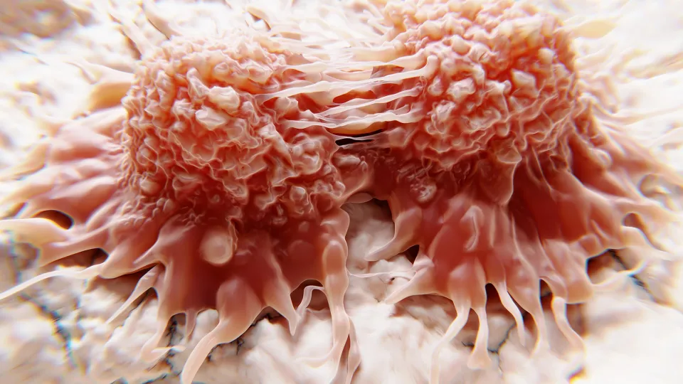
The major challenge faced by physicians is the prevention and treatment of metastasis, the main reason for cancer mortality. Surprisingly, cancer patients presumed cured after primary tumor removal and therapy, can carry non-proliferating ‘dormant’ disseminated cancer cells (DCCs) for years before reactivating to form incurable metastasis. I focused on understanding the biology of dormant DCCs and their reactivation, to target them and prevent relapse. My team led a paradigm shift that is revealing novel cancer biology that diverges from the notion that cancer is perpetually proliferating.
We discovered that a reciprocal crosstalk between DCCs and the microenvironment regulates the inter-conversion between dormancy and proliferation. My lab discovered that an imbalance in p38a/b and ERK1/2 signaling regulates lineage commitment transcription factors (TF) and an epigenetic network that determines dormancy induction (Aguirre-Ghiso et al., JCB 1999, Mol Biol Cell 2001, Oncogene 2002, Liu, Aguirre-Ghiso et al., Cancer Cell, 2002, Aguirre-Ghiso et al., Cancer Res 2004, among others). This work led to identify retinoic acid and TGFb2 in the microenvironment as inducers of the high p38/ERK-signaling ratio, leading to the finding that dormancy (quiescence) is controlled by mechanisms of adult stem cell quiescence (Adam et al., Cancer Res 2009; Bragado et al., Nat Cell Bio, 2013, Sosa et al., Nat Commun 2015; Fluegen et al., & Nat Cell Bio, 2017; Nobre et al., Nature Cancer 2021). These findings have been expanded to reveal that mesenchymal stem cell niches that control hematopoietic stem cell dormancy control dormancy of breast cancer DCCs through a TGFB2 and BMP7-dependent mechanism (Nobre et al.,Nat Cancer, 2021). My lab was one of the first to show that tumor-initiating properties and DCC dormancy are dynamically linked by epigenetic states and a transcriptional program that affects the histone code (Sosa et al., Nat Commun 2015). This work led to an epigenetic strategy to induce dormancy, which is now a clinical trial funded by the V-Foundation and TCI (NCT03572387). We also identified a “dormancy signature” and specific markers enriched in dormant prostate and breast cancer DCCs as well as in the bone marrow of breast cancer patients that predict for prolonged metastasis-free periods (Borgen et al., Breast Cancer Res 2018; Nobre et al., Nature Cancer 2021). We also found how the hypoxic microenvironment in primary tumors primes DCCs epigenetically to become dormant and evade chemotherapy (Fluegen et al., Nat Cell Bio, 2017).
My lab’s work has propelled new questions to the forefront of cancer research, with the unexpected discovery of the mechanisms that allow dormant breast cancer DCCs and metastasis to be initiated very early during cancer evolution disseminating during pre-malignant stages (Harper and Sosa et al., Nature, 2016 and Hosseini et al., Nature, 2016). We further discovered that innate immune cells are key to launch the early dissemination process and the success on metastatic colonization (Linde et al., Nature Commun. 2018). Our program’s impact is further highlighted by the development of successful collaborations with industry that identified pathways (Ranganathan et al., Cancer Ress 2006; 2008; Schewe and Aguirre-Ghiso, PNAS 2008; Cancer Res 2009) and drugs to kill dormant DCCs or induce dormancy of DCCs that are now part of a patent (WO201919111) and developed by a new startup company (HiberCell)(www.hibercell.com), which is conducting clinical trials developed from our findings (NCT04834778, NCT04995094),identification of potential biomarkers and the development of the first clinical trials to induce dormancy of metastatic disease in prostate cancer (NCT03572387).
Selected Publications
Pre-Prints Available Online
1. Deepak K. Singh, Eduardo Farias, Saul Carcamo, Dan Hasson, Dan Sun, Julie Cheung, Ana Rita Nobre, Nupura Kale, Maria Soledad Sosa, Emily Bernstein, Julio A. Aguirre-Ghiso. Epigenetic reprogramming of DCCs into dormancy suppresses metastasis via restored TGFβ–SMAD4 signaling bioRxiv 2021.08.01.454684; doi: https://doi.org/10.1101/2021.08.01.454684
2. Aguirre-Ghiso JA, Nobre AR, Dalla E, et al. A Mesenchymal-like Program of Dormancy controlled by ZFP281 Serves as a Barrier to Metastatic Progression of Early Disseminated Cancer Cells. 18 January 2021, PREPRINT (Version 1) available at Research Square. https://doi.org/10.21203/rs.3.rs-145308/v1
3. Khalil BD, Sanchez R, Rahman T, Rodriguez-Tirado C, Moritsch S, Rodriguez Martinez A, Miles B, Farias E, Mezei M, Cheung JF, Nobre AR, Kale N, Sproll KC, Sosa MS, Aguirre-Ghiso JA. A specific agonist of the orphan nuclear receptor NR2F1 suppresses metastasis through the induction of cancer cell dormancy. bioRxiv(2021).01.30.428967; doi: https://doi.org/10.1101/2021.01.30.428967
4. Borriello L, Coste A, Sharma VP, Karagiannis GS, Lin Y, Wang Y, Ye X, Duran C, Chen X, Dalla E, Singh DK, Oktay MH, Aguirre-Ghiso JA, Condeelis J, Entenberg D. Primary tumor associated macrophages activate programs of invasion and dormancy in disseminating tumor cells. bioRxiv (2021).02.04.429798; doi: https://doi.org/10.1101/2021.02.04.429798
5. Mitchell Fane, Stephen Douglass, Gretchen Alicea, Marie Webster, Vito Rebecca, Gloria Marino, Yash Chhabra, Filipe Almeida, Brett Ecker, Thomas Beer, Hsin-Yao Tang, Andrew Kossenkov, David Speicher, Julio Aguirre-Ghiso, Ashani Weeraratna, Stromal Changes In The Aged Lung Induce An Emergence From Melanoma Dormancy, 16 September (2020), PREPRINT (Version 1) available at Research Square https://doi.org/10.21203/rs.3.rs-61165/v1
Peer Reviewed Original Research Contributions
1. Di Martino J, Nobre AR, Mondal C, Taha I, Farias EF, Fertig E, Naba A, Aguirre-Ghiso JA, Bravo-Cordero JJ. A tumor-derived type III collagen-rich ECM niche regulates tumor cell dormancy, 22 June 2021, PREPRINT (Version 1) available at Research Square [https://doi.org/10.21203/rs.3.rs-580847/v1]. Nature Cancer (2021) (Accepted for Publication)
2. Casanova-Acebes M, Dalla E, Leader A, LeBerichel J, Nikolic J, Morales BM, Brown M, Chang M, Troncoso L, Chen ST, Sastre-Perona A, Park MD, Tabachnikov A, Dhainaut M, Hamon P, Maier B, Sawai CM, Agulló-Pascual E, Schober M, Brown B, Reizis B, Marron T, Kenigsberg E, Moussion C, Benaroch P, Aguirre-Ghiso JA, Merad M. Lineage tracing reveals the unique pro-tumorigenic niche role of tissue-resident macrophages in early cancer lesions. Nature 595, 578–584 (2021). In the top 5% of all research outputs scored by Altmetric
3. Nobre R, Risson E, Singh DK, Di Martino JS, Cheung JF, Wang J, Johnson J, Russnes HG, Bravo-Cordero JJ, Birbrair A, Naume B, Azhar M, Frenette PS, Aguirre-Ghiso JA. Bone marrow NG2+/Nestin+ mesenchymal stem cells drive DTC dormancy via TGF-β2. Nature Cancer 2, 327–339 (2021). https://doi.org/10.1038/s43018-021-00179-8 In the top 5% of all research outputs scored by Altmetric
4. Lam T, Aguirre-Ghiso JA, Geller MA, Aksan A, Azarin SM. Immobilization rapidly selects for chemoresistant ovarian cancer cells with enhanced ability to enter dormancy. Biotechnol Bioeng. (2020) Oct;117(10):3066-3080. doi: 10.1002/bit.27479. Epub 2020 Jul 9.
5. Teh JL, Purwin TJ, Han A, Chua V, Patel P, Baqai U, Liao C, Bechtel N, Sato T, Davies MA, Aguirre-Ghiso JA, Aplin AE. Metabolic adaptations to MEK and CDK4/6 co-targeting in uveal melanoma. Mol Cancer Ther. (2020) May 19: molcanther.1016.2019. doi: 10.1158/1535-7163.MCT-19-1016. Epub ahead of print. PMID: 32430489.
6. Liu PH, Shah RB, Li Y, Arora A, Ung PM, Raman R, Gorbatenko A, Kozono S, Zhou XZ, Brechin V, Barbaro JM, Thompson R, White RM, Aguirre-Ghiso JA, Heymach JV, Lu KP, Silva JM, Panageas KS, Schlessinger A, Maki RG, Skinner HD, de Stanchina E, Sidi S. An IRAK1-PIN1 signalling axis drives intrinsic tumour resistance to radiation therapy. Nat Cell Biol. (2019) Feb;21(2):203-213. doi: 10.1038/s41556-018-0260-7. Epub 2019 Jan 21.
7. Lapadula D, Farias E, Randolph CE, Purwin TJ, McGrath D, Charpentier TH, Zhang L, Wu S, Terai M, Sato T, Tall GG, Zhou N, Wedegaertner PB, Aplin AE, Aguirre-Ghiso JA, Benovic JL. Effects of Oncogenic Gαq and Gα11 Inhibition by FR900359 in Uveal Melanoma. Mol Cancer Res. (2019) Apr;17(4):963-973. doi: 10.1158/1541-7786.MCR-18-0574. Epub 2018 Dec 19. In the top 5% of all research outputs scored by Altmetric and picked up by 16 news stories from 15 outlets.
8. Borgen E, Rypdal MC, Sosa MS, Renolen A, Schlichting E, Lønning PE, Synnestvedt M, Aguirre-Ghiso JA, Naume B. NR2F1 stratifies dormant disseminated tumor cells in breast cancer patients. Breast Cancer Research(2018) 20 (1), 120. In the top 5% of all research outputs scored by Altmetric and picked up by 9 news stories from 9 outlets.
9. Linde N, Casanova-Acebes M, Sosa MS, Mortha A, Rahman A, Farias EF, Harper K, Tardio E, Reyes Torres I, Jones J, Condeelis J, Merad M, and Aguirre-Ghiso JA.
Macrophages orchestrate breast cancer early dissemination and metastasis. Nature Communications 9, 21(2018) doi:10.1038/s41467-017-02481-5. In the top 5% of all research outputs scored by Altmetric. Highlighted in Faculty of 1000 Prime: El-Deiry W and Zhao S: F1000Prime Recommendation of [Linde N et al., Nat Commun 2018 9(1):21]. In F1000Prime, 11 Jan 2018; 10.3410/f.732394311.793541160. Highlighted in Science Translational Medicine:
https://stm.sciencemag.org/content/10/425/eaar7521?intcmp=trendmd-stm
10. Entenberg D, Pastoriza JM, Oktay MH, Voiculescu S, Wang Y, Sosa MS, Aguirre-Ghiso JA, Condeelis J. Time-lapsed, large-volume, high-resolution intravital imaging for tissue-wide analysis of single cell dynamics. Methods(2017) Sep 1; 128:65-77. doi: 10.1016/j.ymeth.2017.07.019.
11. Fluegen G, Avivar-Valderas A, WangY, PadgenM, WilliamsJK, Verkhusha V, Cheung JF, Entenberg D, Castracane J, Keely PJ, Condeelis J, Aguirre-Ghiso JA. Phenotypic heterogeneity of disseminated tumor cells is predetermined by primary tumor hypoxic microenvironments. Nature Cell Biology (2017) doi:10.1038/ncb3465.Cover Article. Highlighted in Science Translational Medicine Editor’s Choice, Science Translational Medicine 15 Feb 2017: Vol. 9, Issue 377, eaam6063 DOI: 10.1126/scitranslmed.aam6063
12. Harper K, Sosa MS, Hosseini H, Entenberg D, Avivar-Valderas A, Nagi C, Davis RJ, Farias EF, Condeelis J, Klein C, Aguirre-Ghiso JA. Mechanism of early dissemination and metastasis during early stages of HER2+ mammary cancer. Nature (2016) Dec 14. doi: 10.1038/nature20785. [Epub ahead of print] PMID:27974799. Paper highlighted in Nature News and Views. Nature. 2016 Dec 14. doi: 10.1038/nature21104. Highlighted by Faculty of 1000. https://f1000.com/prime/727099751; Highlighted in Nature Reviews Cancer http://www.nature.com/nrc/journal/v17/n2/full/nrc.2017.4.html
13. Hosseini, H, Harper K, Obradovic M, Sosa MS, Nanduri LK, Werno C, Hoffman, M, Ehrl C, Maneck M, Patwary N, Haunschild G, Reimelt C, Weeber F, Hartkopf A, Taran FA, Brucker SY, Fehm T, Meister G, Aguirre-Ghiso JA, Klein CA. Early dissemination seeds metastasis in breast cancer. Nature. (2016) Dec 14. doi: 10.1038/nature20609. [Epub ahead of print] PMID:27974798. Paper highlighted in Nature News and Views. Nature. 2016 Dec 14. doi: 10.1038/nature21104. Highlighted in Nature Reviews Cancer http://www.nature.com/nrc/journal/v17/n2/full/nrc.2017.4.html
14. Yumoto K, Eber MR, Wang J, Cackowski FC, Decker AM, Lee E, Nobre AR, Aguirre-Ghiso JA, Jung Y, Taichman RS. Axl is required for TGF-β2-induced dormancy of prostate cancer cells in the bone marrow. Scientific Reports (2016) Nov 7; 6:36520. doi: 10.1038/srep36520.
15. Morris BA, Burkel B, Ponik SM, Fan J, Condeelis JS, Aguire-Ghiso JA, Castracane J, Denu JM, Keely PJ. Collagen Matrix Density Drives the Metabolic Shift in Breast Cancer Cells. EBioMedicine (2016) Nov; 13:146-156. doi: 10.1016/j.ebiom.2016.10.012. PubMed PMID: 27743905.
16. Cackowski FC, Eber MR, Rhee J, Decker A, Yumoto K, Berry JE, Lee E, Shiozawa Y, Jung Y, Aguirre-Ghiso JA,Taichman RS. Mer Tyrosine Kinase Regulates Disseminated Prostate Cancer Cellular Dormancy. J Cell Biochem(2016) Oct 18. doi: 10.1002/jcb.25768. PMID: 27753136
17. Chung CY, Sun Z, Mullokandov G, Bosch A, Qadeer ZA, Cihan E, Rapp Z, Parsons R, Aguirre-Ghiso JA, Farias EF, Brown BD, Gaspar-Maia A, Bernstein E. Cbx8 acts non-canonically in breast cancer to promote Notch signaling. Cell Reports (2016) Jul 12;16(2):472-86. doi: 10.1016/j.celrep.2016.06.002. Epub 2016 Jun 23. PMID: 27346354
18. Williams JK, Entenberg D, Wang Y, Avivar-Valderas A, Padgen M, Clark A, Aguirre-Ghiso JA, Castracane J, Condeelis JS. Validation of a device for the active manipulation of the tumor microenvironment during intravital imaging. Intravital. (2016)
19. Szulczewski JM, Inman DR, Entenberg D, Aguirre-Ghiso JA, Castracane J, Condeelis J, Eliceiri KW, Keely PJ. In Vivo Visualization of Stromal Macrophages via label-free FLIM-based metabolite imaging. Scientific Reports(2016) May 25; 6:25086. doi: 10.1038/srep25086. PMID: 27220760
20. Li M, Pathak RR, Lopez-Rivera E, Friedman SL, Aguirre-Ghiso JA, Sikora AG. The In Ovo Chick Chorioallantoic Membrane (CAM) Assay as an Efficient Xenograft Model of Hepatocellular Carcinoma. J Vis Exp. (2015) Oct 9; (104). doi: 10.3791/52411.
21. Alsadeq A, Strube S, Krause S, Carlet M, Jeremias I, Vokuhl C, Loges S, Aguirre-Ghiso JA, Trauzold A, Cario G, Stanulla M, Schrappe M, Schewe DM. Effects of p38α/β inhibition on acute lymphoblastic leukemia proliferation and survival in vivo. Leukemia. (2015) Jun 24. doi: 10.1038/leu.2015.153. [Epub ahead of print] PMID: 26104660
Adomako A, Calvo V, Biran N, Osman K, Chari A, Paton JC, Paton AW, Moore K, Schewe DM, Aguirre-GhisoJA. Identification of markers that functionally define a quiescent multiple myeloma cell sub-population surviving bortezomib treatment. BMC Cancer (2015) May 30; 15:444. doi: 10.1186/s12885-015-1460-1. PMCID: PMC4448210. Highlighted by BMC Cancer in the “Highly Accessed” category.
22. Hasegawa D, Calvo V, Avivar-Valderas A, Lade A, Chou HI, Lee YA, Farias EF, Aguirre-Ghiso JA, Friedman SL. Epithelial Xbp1 is Required for Cellular Proliferation and Differentiation During Mammary Gland Development. Mol Cell Biol. (2015) Feb 23. pii: MCB.00136-15. PMID: 25713103. Cover article for the Feb 23 issue of Mol Cell Biol.
23. Sosa MS, Parikh F, Maia AG, Estrada Y, Bosch A, Bragado P, Ekpin E, George A, Zheng Y, Lam HM, Morrissey C, Chung CY, Farias EF, Bernstein E, Aguirre-Ghiso JA. NR2F1 controls tumour cell dormancy via SOX9- and RARβ-driven quiescence programmes. Nat Commun (2015) Jan 30; 6:6170. doi: 10.1038/ncomms7170. PubMed PMID:25636082; PubMed Central PMCID: PMC4313575. In the top 10% of all articles ever tracked by Altmetric.
24. Chéry L, Lam HM, Coleman I, Lakely B, Coleman R, Larson S, Aguirre-Ghiso JA, Xia J, Gulati R, Nelson PS, Montgomery B, Lange P, Snyder LA, Vessella RL, Morrissey C. Characterization of single disseminated prostate cancer cells Reveals tumor cell heterogeneity and identifies dormancy associated pathways. Oncotarget (2014) Oct 30;5(20):9939-51. PubMed PMID: 25301725; PubMed Central PMCID: PMC4259449.
25. Lopez-Rivera E, Jayaraman P, Parikh F, Davies MA, Ekmekcioglu S, Izadmehr S, Milton DR, Chipuk JE, Grimm EA, Estrada Y, Aguirre-Ghiso JA, Sikora AG. Inducible nitric oxide synthase drives mTOR pathway activation and proliferation of human melanoma by reversible nitrosylation of TSC2.Cancer Res. (2014) Feb 15;74(4):1067-78. doi: 10.1158/0008-5472.CAN-13-0588. Epub 2014 Jan 7. PMID: 24398473 [PubMed - indexed for MEDLINE]
26. Bragado P, Estrada Y, Parikh F, Krause S, Capobianco C, Farina HG, Schewe DM, Aguirre-Ghiso JA. TGFβ2 dictates disseminated tumour cell fate in target organs through TGFβ-RIII and p38α/β signalling. Nat. Cell. Bio. (2013).Nov;15(11):1351-61. doi: 10.1038/ncb2861. Epub 2013 Oct 27. PMID: 24161934;PMCID: not available. Featured in Nat Cell Bio Cover same issue. Highlighted in Science Signaling Editor’s Choice, Cancer Research Breaking Advances section and several public news outlets. Featured in the Cover of the November 15th issue of Nat Cell Bio. Ranks #6 for articles of similar age in Nat Rev Cancer and is in the top 5% of all articles ever tracked by Altmetric.
27. Begley U, Sosa MS, Avivar-Valderas A, Patil A, Endres L, Estrada Y, Chan CTY, Su D, Dedon PC, Aguirre-Ghiso JA, Begley T. A human tRNAmethyltransferase 9-like protein prevents tumor growth by regulating LIN9 and the hypoxic response. EMBO Mol Med (2013) Feb 4. doi: 10.1002/emmm.201201161.
28. Avivar-Valderas A, Bobrovnikova-Marjon E, Diehl JA, Bardeesy N, Debnath J, Aguirre-Ghiso JA. Regulation of autophagy during ECM detachment is linked to a selective inhibition of mTORC1 by PERK. Oncogene(2012), Nov 19. doi: 10.1038/onc.2012.512.
29. Kim RS, Avivar-Valderas A, Estrada A, Bragado P, Sosa MS, Aguirre-Ghiso JA, Segall JE. Dormancy signatures and metastasis in estrogen receptor positive breast cancer. PLoS One (2012); 7(4):e35569. Epub 2012 Apr 18.
30. Bragado P, Estrada Y, Sosa MS, Avivar-Valderas A, Cannan D, Genden E, Teng M, Ranganathan AC, Wen HC, Kapoor A, Bernstein E, Aguirre-Ghiso JA. Analysis of marker-defined HNSCC subpopulations reveals a dynamic regulation of tumor initiating properties. PLoS One. (2012); 7(1):e29974. PMCID: PMC3262798.
31. Tong CC, Lau KH, Rivera M, Cannan D, Aguirre-Ghiso JA, Sikora AG, Gupta V, Forsythe K, Ko EC, Misiukiewicz K, Gurudutt V, Teng MS, Packer SH, Genden EM, Kao J. Prognostic significance of p16 in locoregionally advanced head and neck cancer treated with concurrent 5-fluorouracil, hydroxyurea, cetuximab and intensity-modulated radiation therapy. Oncol Rep. (2012) May; 27(5):1580-6. doi: 10.3892/or.2012.1679.
32. Gutiérrez-Uzquiza Á, Arechederra M, Bragado P, Aguirre-Ghiso JA, Porras A. p38a mediates cell survival in response to oxidative stress via induction of antioxidant genes. Effect on p70S6K pathway. J Biol Chem. (2012)Jan 20;287(4):2632-42. Epub 2011 Dec 2.
33. Fu S, Rivera M, Ko EC, Sikora AG, Chen CT, Vu HL, Cannan D, Eisenstein S, Rosenstein BS, Aguirre-Ghiso JA, Chen SH, Kao J. Combined Inhibition of Epidermal Growth Factor Receptor and Cyclooxygenase-2 as a Novel Approach to Enhance Radiotherapy. J Cell SciTher (2011) S1:002. doi:10.4172/2157-7013.S1-002.
34. Avivar-Valderas A, Salas E, Bobrovnikova-Marjon E, Diehl JS, Nagi C, Debnath J, Aguirre-Ghiso JA, PERK integrates autophagy and oxidative stress responses to promote survival during ECM detachment. Mol Cell Biol. (2011) Sep; 31(17):3616-29. PMC3165554 (Cover article)
35. Wen HC, Avivar-Valderas A, Sosa MS, Girinius N, Farias EF, Davis R, Aguirre-Ghiso JA. p38 signaling regulates ATF2/c-Jun and BimEL to induce anoikis and lumen formation during mammary morphogenesis. Science Signaling (2011) 4 (174), ra34. [DOI: 10.1126/scisignal.2001684].http://stke.sciencemag.org/cgi/content/abstract/sigtrans;4/174/ra34
36. Ishii Y, Papa Y, Bahadur U, Yue Z, Aguirre-Ghiso JA, Shioda T, Waxman S, Germain D. Bortezomib enhances the efficacy of fulvestrant by amplifying the aggregation of theestrogen receptor, which leads to a pro-apoptotic unfolded protein response. Clin. Cancer Res. (2011) Feb 3.
37. Sequeira SJ, Wen HC, Avivar-Valderas A, Aguirre-Ghiso JA. Inhibition of eIF2a dephosphorylation inhibits ErbB2-induced deregulation of mammary acinar morphogenesis.BMC Cell Biology. (2009) Sep 15;10:64.
38. Schewe DM. Aguirre-Ghiso JA. Inhibition of eIF2 dephosphorylation eliminates quiescent multiple myeloma cells surviving proteasome inhibitor therapy. Cancer Res. (2009) Feb 15;69(4):1545-52.
39. Adam AP, George A, Bragado P, Iglesias BV, Schewe D, Ranganathan A, Kourtidis A, Conklin DS, Aguirre-Ghiso JA. Computational identification of a p38SAPK regulated transcription factor network required for tumor cell quiescence. Cancer Res. (2009) Jul 15; 69(14):5664-72. Epub 2009 Jul 7.
40. Schewe DM, Aguirre-Ghiso JA. ATF6α-Rheb-mTOR signaling promotes survival of dormant tumor cells in vivo. Proc Natl Acad Sci USA (2008) Jul 29;105(30):10519-24. Highlighted in “This week in PNAS” section of the journal. http://www.pnas.org/content/105/30/10271.full?sid=79541a8a-11a2-41cc-aeef- Faculty of 1000 evaluation: www.f1000biology.com/article/id/1119501/evaluation
41. Ranganathan AC, Ohja S, Kourtidis A, Conklin D, Aguirre-Ghiso JA. Dual function of PERK in tumor cell growth arrest and survival. Cancer Res (2008); 68(9): 1–9.
42. Sequeira SJ, Ranganathan AC, Adam AP, Iglesias BV, Farias EF, Aguirre-Ghiso JA. Inhibition of Proliferation by PERK Regulates Mammary Acinar Morphogenesis and Tumor Formation. PLoS ONE. (2007) Jul 18; 2:e615.
43. Baron TE, Lastro MT, Ranganathan AC, Tenenbaum SA, Conklin DS, Aguirre-Ghiso JA. Ribonomic and Short Hairpin RNA Gene Silencing Methods to Explore Functional Gene Programs Associated with Tumor Growth Arrest. Methods Mol Biol. (2007); 383:227-44.
44. Shinohara M, Mikhailov AV, Aguirre-Ghiso JA, Rieder CL. ERK1/2 activation is required for timely progress through early G2 but it is not directly involved in the G2/M or M/A transitions in mammalian cells. Molecular Biology of the Cell. (2006) Dec 1st.17: 5227-5240.
45. Chaurasia P, Aguirre-Ghiso JA, Liang OD, Gardsvoll H, Ploug M, Ossowski LA. Region in uPAR domain III controlling a functional association with a5b1 integrin and tumor growth. J. Biol. Chem. (2006) May 26; 281(21): 14852-63.
46. Ranganathan AC, Zhang L, Adam AP, Aguirre-Ghiso JA. Functional coupling of p38-induced upregulation of BiP and activation of RNA-dependent protein kinase–like ER kinase (PERK) to drug resistance of dormant carcinoma cells. Cancer Research (2006); 66: (3) 1702-11.
47. Connor KM, Subbaram S, Regan KJ, Nelson KK, Mazurkiewicz JE, Bartholomew PJ, Aplin AE, Tai YT, Aguirre-Ghiso JA, Flores SC, Melendez JA. Mitochondrial H2O2 Regulates the Angiogenic Phenotype Via PTEN Oxidation. J. Biol. Chem. (2005) 29; 280(17): 16916-24.
48. Aguirre-Ghiso JA, Ossowski L, Rosenbaum SK. GFP tagging of ERK and p38 pathways reveals novel dynamics of pathway activation during primary and metastatic growth. Cancer Res (2004); 64(20): 7336-45.Faculty of 1000 evaluation at: www.f1000biology.com/article/15492254/evaluation
49. Mazzoni E, Adam A, Bal de Kier Joffe E, Aguirre-Ghiso JA. Immortalized mammary epithelial cells overexpressing PKC acquire a malignant phenotype and become tumorigenic in vivo. Molecular Cancer Res. (2003) 1:776-787
50. Aguirre-Ghiso JA, Estrada Y, Liu D, Ossowski L. ERK (MAPK) activity as a determinant of tumor growth and dormancy; regulation by p38 (SAPK). Cancer Res. (2003); 63(7): 1684-95.
51. Puricelli L, Proiettii CJ, Labriola L, Salatino M, Balana ME, Aguirre-Ghiso JA, Lupu R, Pignataro OP, Charreau EH, Bal de Kier Joffe E, Elizalde PV. Heregulin inhibits proliferation via ERKs and phosphatidyl-inositol 3-kinase activation but regulates urokinase plasminogen activator independently of these pathways in metastatic mammary tumor cells. Int. J. Cancer. (2002) 100(6): 642-53.
52. Liu D, Aguirre-Ghiso JA, Estrada Y, Ossowski L. EGFR is a transducer of the urokinase receptor initiated signal that is required for in vivo growth of a human carcinoma. Cancer Cell (2002); 1: 445-457(Cover). Preview in the same issue: http://www.cell.com/cancer-cell/abstract/S1535-6108(02)00076-4 Faculty of 1000 Evaluation at: www.f1000biology.com/article/12124174/evaluation
53. Aguirre-Ghiso JA. Inhibition of FAK signaling activated by urokinase receptor induces human carcinoma dormancy in vivo. Oncogene (2002) 21(16): 2513-24.
54. Ladeda V, Frankel P, Feig LA, Foster DA, Bal de Kier Joffe E, Aguirre-Ghiso JA. RalA mediates v-Src, v-Ras, and v-Raf regulation of CD44 and fibronectin expression in NIH3T3 fibroblasts. BiochemBiophys Res Commun. (2001) 283(4): 854-61.
55. Aguirre-Ghiso JA, Liu D, Mignatti D, Kovalski K, Ossowski L. Urokinase receptor and fibronectin regulate the ERKMAPK to p38MAPK activity ratios that determine carcinoma cell proliferation or dormancy in vivo. Mol. Biol. Cell (2001). 12, 4, 863-879.
56. Aguirre-Ghiso JA, Kovalski K, Ossowski L. Tumor dormancy due to urokinase receptor down-regulation in human carcinoma involves integrin and MAPK signaling. J. Cell Biol. (1999) 147, 89-103. Highlighted in the In Brief section of JCB.
57. Aguirre-Ghiso JA, Frankel P, Farías E, Lu Z, Jiang H, Olsen A, Feig L, Bal de Kier Joffé E, Foster D. Ral-A requirement for v-Src- and v-Ras-induced tumorigenicity and overproduction of urokinase-type plasminogen activator. Involvement of metalloproteinases. Oncogene (1999), 18, 4718-4725.
58. Urtreger AJ, Aguirre-Ghiso JA, Werbahj S, Puricelli L, Muro A, Bal de Kier Joffé E. Involvement of fibronectin in the regulation of urokinase production and binding in murine mammary tumor cells.Int. J. Cancer, (1999), vol. 82, 748-753.
59. Ladeda V, Aguirre-Ghiso JA, Bal de Kier Joffé E. Function and Expression of CD44 During Spreading and Migration of Murine Carcinoma cells. Exp. Cell Res. (1998); 242:515-527.
60. Aguirre-Ghiso JA, Farías EF, Alonso DF, Bal de Kier Joffé E. Secretion of urokinase and metalloproteinase-9 induced by staurosporine is dependent on a tyrosine-kinase pathway in mammary tumor cells. Int. J. Cancer (1998) 76: 362-367.
61. Farías EF, Aguirre-Ghiso JA, Ladeda V, Bal de Kier Joffé E. Verapamil Inhibits Proteases Production, Local Invasion and Metastasis Development of Murine Carcinoma Cells. Int. J. Cancer. (1998) 78 (6): 727-734.
62. Aguirre-Ghiso JA, Farías EF, Alonso DF, Arregui C, Bal de Kier Joffé E E. A Phospholipase D and Protein Kinase C Inhibitor Blocks the Spreading of Murine Mammary Adenocarcinoma Cells Altering F-Actin and Integrin Point Contact Distribution. Int. J. Cancer. (1997) 71: 881-890.
63. Aguirre-Ghiso JA, Alonso DF, Farías E, Bal de Kier Joffé E. Overproduction of urokinase-type plasminogen activator is regulated by phospholipase D and protein kinase C pathways in murine mammary adenocarcinoma cells. Biochem. Biophys. Acta (1997) 1356: 171-184.
64. Aguirre-Ghiso JA, Diament M, D'elia I, Bal de Kier Joffé E, Klein S. Effect of in vivo culture of murine mammary adenocarcinoma cells on tumor and metastatic growth. Tumor Biol. (1997); 18: 41-52.
65. Aguirre-Ghiso JA, Farías E, Fernandez DM, Alonso DF, Bal de Kier Joffé E. Down modulation of tumor cells-associated proteolytic activity by n-butanol treatment in cultured murine mammary adenocarcinoma cells. Int. J Oncol. (1996); 8: 35-39.
66. Klein S, Jasnis MA, Diament M, Davel L, Aguirre Ghiso JA, de Bonaparte YP. Immunomodulation by soluble factors from tumor cells cultured in vivo in diffusion chambers. Tumor Biol. (1994);15: 160-165.
Reviews, Commentaries, Perspectives and other Peer Reviewed Publications
1. Aguirre-Ghiso JA. Translating the Science of Cancer Dormancy to the Clinic. Cancer Res (2021)August 24. DOI: 10.1158/0008-5472.CAN-21-1407
2. Atkins MB, Curiel-Lewandrowski C, Fisher DE, Swetter SM, Tsao H, Aguirre-Ghiso JA, Soengas MS, Weeraratna AT, Flaherty KT, Herlyn M, Sosman JA, Tawbi HA, Pavlick AC, Cassidy PB, Chandra S, Chapman PB, Daud A, Eroglu Z, Ferris LK, Fox BA, Gerhsenwald JE, Gibney GT, Grossman D, Hanks BA, Hanniford D, Hernando E, Jeter JM, Johnson DB, Khleif SN, Kirkwood JM, Leachman SA, Mays D, Nelson KC, Sondak VK, Sullivan RJ, Merlino G. The State of Melanoma: Emergent Challenges and Opportunities. Clin Cancer Res. (2021) Jan 7:clincanres.4092.2020. doi: 10.1158/1078-0432.CCR-20-4092.
3. Risson E, Nobre AR, Maguer-Satta V, Aguirre-Ghiso JA. The current paradigm and challenges ahead for the dormancy of disseminated tumor cells. Nat Cancer (2020). https://doi.org/10.1038/s43018-020-0088-5. In the top 5% of all research outputs scored by Altmetric
4. Vabret N, Britton GJ, Gruber C, Hegde S, Kim J, Kuksin M, Levantovsky R, Malle L, Moreira A, Park MD, Pia L, Risson E, Saffern M, Salomé B, Esai Selvan M, Spindler MP, Tan J, van der Heide V, Gregory JK, Alexandropoulos K, Bhardwaj N, Brown BD, Greenbaum B, Gümüş ZH, Homann D, Horowitz A, Kamphorst AO, Curotto de Lafaille MA, Mehandru S, Merad M, Samstein RM; Sinai Immunology Review Project. Immunology of COVID-19: Current State of the Science. Immunity. (2020) Jun 16;52(6):910-941. doi: 10.1016/j.immuni.2020.05.002. Epub 2020 May 6.
5. Zijlstra A, Von Lersner A, Yu D, Borrello L, Oudin M, Kang Y, Sahai E, Fingleton B, Stein U, Cox TR, Price JT, Kato Y, Welm AL, Aguirre-Ghiso JA; Board Members of the Metastasis Research Society. The importance of developing therapies targeting the biological spectrum of metastatic disease. Clin. Exp. Metastasis. (2019) Aug;36(4):305-309. doi: 10.1007/s10585-019-09972-3. Epub 2019 May 17.
6. Aguirre-Ghiso JA. How dormant cancer persists and reawakens. Science. (2018) Sep 28;361(6409):1314-1315. doi: 10.1126/science.aav0191.
7. Aguirre-Ghiso JA, MS Sosa. Emerging topics on disseminated cancer cell dormancy and the paradigm of metastasis. Annual Review of Cancer Biology (2018), 2, 377-393
8. Nobre AR, Entenberg D, Wang Y, Condeelis J, Aguirre-Ghiso JA. The Different Routes to Metastasis via Hypoxia-Regulated Programs. Trends Cell Biol. (2018) Jul 21. pii: S0962-8924(18)30109-0. doi: 10.1016/j.tcb.2018.06.008. [Epub ahead of print] Review. PMID: 30041830
9. Fingleton B, Lange K, Caldwell B, Bankaitis KV; Board of the Metastasis Research Society. Perspective on the interpretation of research and translation to clinical care with therapy-associated metastatic breast cancer progression as an example. ClinExp Metastasis. (2017) Dec;34(8):443-447. doi: 10.1007/s10585-017-9872-8. Epub 2018 Feb 26 PMID: 29484519
10. Linde N, Fluegen G, Aguirre-Ghiso JA. The Relationship Between Dormant Cancer Cells and Their Microenvironment. Adv Cancer Res. (2016); 132:45-71. doi: 10.1016/bs.acr.2016.07.002. Epub 2016 Aug 25. PMID:27613129.
11. Klionsky DJ, Abdelmohsen K, Abe A, Abedin MJ, Abeliovich H, Acevedo Arozena A, Adachi H, Adams CM, Adams PD, Adeli K, Adhihetty PJ, Adler SG, Agam G, Agarwal R, Aghi MK, Agnello M, Agostinis P, Aguilar PV, Aguirre-Ghiso JA, et al. Guidelines for the use and interpretation of assays for monitoring autophagy. Autophagy (2016) Jan 2;12(1):1-222. No abstract available. PMID: 26799652
12. Goossens N, Hoshida Y, Aguirre-Ghiso JA. Origin and interpretation of cancer transcriptome profiling: the essential role of the stroma in determining prognosis and drug resistance. EMBO Mol Med (2015) Aug 3. pii: e201505284. doi: 10.15252/emmm.201505284. [Epub ahead of print]
13. Sosa MS, Bragado P, Aguirre-Ghiso JA. Mechanisms of disseminated cancer cell
dormancy: an awakening field. Nat Rev Cancer (2014) Sep;14(9):611-22. doi:
10.1038/nrc3793. Epub 2014 Aug 14. Review. PubMed PMID: 25118602; PubMed Central
PMCID: PMC4230700.
-Cover article for the September 14th issue of Nat Rev Cancer
-Ranks 6 for articles of similar age in Nat Rev Cancer and is in the top 5% of all articles ever tracked by Altmetric.
14. Linde N, Sosa MS, Aguirre-Ghiso JA. Integration of microenvironmental and stress signaling antagonizes colorectal cancer progression. EMBO J. (2014) Aug 18;33(16):1737-9. doi: 10.15252/embj.201489364. Epub 2014 Jul 14. PubMed PMID: 25024435; PubMed Central PMCID: PMC4195757.
15. Avivar-Valderas A, Wen HC, Aguirre-Ghiso JA. Stress signaling and the shaping
of the mammary tissue in development and cancer. Oncogene (2014) Nov
27;33(48):5483-90. doi: 10.1038/onc.2013.554. Epub 2014 Jan 13. Review. PubMed
PMID: 24413078; PubMed Central PMCID: PMC4096615.
16. Aguirre-Ghiso, JA, Bragado P, Sosa MS. Metastasis awakening: targeting dormant cancer. Nat Med. (2013) Mar;19(3):276-7. doi: 10.1038/nm.3120. PMID: 23467238 [PubMed - indexed for MEDLINE] PMCID: PMC3651698
17. Sosa MS, Bragado P, Debnath J, Aguirre-Ghiso JA. Regulation of tumor cell dormancy by tissue microenvironments and autophagy. AdvExp Med Biol. (2013);734:73-89. doi: 10.1007/978-1-4614-1445-2_5.
18. Klionsky DJ, Abdalla FC, Abeliovich H, Abraham RT, Acevedo-Arozena A, Adeli K, Agholme L, Agnello M, Agostinis P, Aguirre-Ghiso JA, et al., Guidelines for the use and interpretation of assays for monitoring autophagy. Autophagy (2012) Apr;8(4):445-544.
19. Bragado P, Sosa MS, Keely P, Condeelis J, Aguirre-Ghiso JA. Microenvironments dictating tumor cell dormancy. Recent Results Cancer Res. (2012); 195:25-39.
20. Cagan RL, Aguirre-Ghiso JA. A local view of cancer. Dev Cell. (2012) Mar 13;22(3):472-4.
21. Genden EM, Aguirre-Ghiso JA. Oropharyngeal cancer biology and treatment: insights from messenger RNA sequence analysis and transoral robotic surgery. Mayo Clin Proc. (2012) Mar;87(3):211-2.
22. Sosa MS, Avivar-Valderas A, Bragado P, Wen HC, Aguirre-Ghiso JA. Molecular Pathways: ERK1/2 and p38α/β signaling in tumor cell quiescence: opportunities to control dormant residual disease. Invited Article. Clin. Cancer Res. (2011) Sep 15;17(18):5850-7.
23. Prince A, Aguirre-Ghiso JA, Genden E, Posner M, Sikora A. Head and neck squamous cell carcinoma: new translational therapies. Mt. Sinai J. Med. (2010). 77 (6): 684-99.
24. Aguirre-Ghiso JA. On the theory of tumor self-seeding: implications for metastasis progression in humans. Breast Cancer Res. (2010) Apr 28;12(2):304. Viewpoint
25. Ossowski L, Aguirre-Ghiso JA. Dormancy of metastatic melanoma. Pigment Cell Melanoma Res. (2010)Feb;23(1):41-56. Epub 2009 Oct 19.
26. Allgayer H, Aguirre-Ghiso JA. The urokinase receptor (u-PAR) - a link between tumor cell dormancy and minimal residual disease in bone marrow? APMIS (2008);116 (7-8) 602-614.
27. Aguirre-Ghiso JA. Models, mechanisms and clinical evidence for cancer dormancy. Nature Reviews Cancer. (2007). Nov; 7(11): 834-46. Invited peer reviewed article (Cover and featured article). http://www.nature.com/nrc/journal/v7/n11/index.html
28. Aguirre-Ghiso JA. The problem of cancer dormancy: understanding the basic mechanisms and identifying therapeutic opportunities. Cell Cycle. (2006) Aug; 5(16): 1740-3. (Guest Editor). Introductory Review. Invited peer reviewed article. Cover.
29. Ranganathan AC, Adam AP and Aguirre-Ghiso JA. Opposing roles of mitogenic and stress signaling pathways in the induction of cancer dormancy. Cell Cycle. (2006) Aug; 5(16): 1799-807. Cover.
30. Ranganathan AC, Adam AP, Zhang L, and Aguirre-Ghiso JA. Tumor cell dormancy induced by p38SAPK and ER-stress signaling; an adaptive advantage for metastatic cells? Cancer Biology & Therapy (2006) Jul; 5(7): 729-35.
31. Tenenbaum SA, Aguirre-Ghiso JA. Dephosphorylation Shows SR Proteins the Way Out. Molecular Cell (2005); 20 (4); 499-501.
32. Ossowski L, Aguirre-Ghiso JA. Urokinase receptor and integrin partnership: coordination of signaling for cell adhesion, migration and growth. Current Opinion in Cell Biology (2000) 12(5): 613-20.
33. Bal de Kier Joffe E, Mazzoni EO, Aguirre-Ghiso JA. Signaling pathways regulating the expression of proteases during tumor progression. (In Spanish) Medicina (B Aires) (2000); 60:34-40.
34. Ossowski L, Aguirre-Ghiso JA, Liu D, Yu W, Kovalski K. The role of plasminogen activator receptor in cancer invasion and dormancy. Medicina (Buenos Aires) (1999) 59, 547-552.
35. Aguirre-Ghiso JA, Alonso DF, Farías EF, Gomez D, Bal de Kier Joffé E. Deregulation of the signaling pathways controlling urokinase production. Its relationship with the invasive phenotype. Eur. J. Biochem.(1999) 263, 2, 295-304 (Cover).
Books and Book Chapters
- Sosa MS, Bragado P, Debnath J, Aguirre-Ghiso JA. Regulation of Tumor Cell Dormancy by Tissue Microenvironments and Autophagy. Systems Biology of Tumor Dormancy. Editors, Enderling H, Almog N and Hlatky L. (2012) Springer Science + Business Media, INC.http://www.springerlink.com/content/u44l178561014r11/
- Bragado P, Sosa MS, Keely P, Condeelis J, Aguirre-Ghiso JA. Microenvironments dictating tumor cell dormancy. Minimal Residual Disease and Circulating Tumor Cells in Breast Cancer. (Eds: K. Pantel, C. Sotiriou, M. Ignatiadis). Recent Results Cancer Res. (2012); 195:25-39. doi: 10.1007/978-3-642-28160-0_3.
- Aguirre-Ghiso JA, Alonso DF, Farias EF. Function and expression of the uPA/uPAR system in cancer metastasis. Cancer Metastasis: Biologic Basis and Therapeutics. Cambridge University Press, Editors: Danny Welch, Danny R. Welch, Ph.D., David C. Lyden, MD, PhD, and Beth Psaila, MD. (2009).http://www.springer.com/biomed/cancer/book/978-1-4419-6614-8
- Bragado P, Ranganathan AC, Aguirre-Ghiso JA. Dormancy of Disseminated Tumor Cells: Reciprocal crosstalk with the microenvironment. The Tumor Microenvironment-Cancer Drug Discovery and Development. Springer Science + Business Media LLC, Humana Press. Dr. Rebecca Bagley, Editor. (2010), Part 2, pp229-254. http://www.cambridge.org/gb/knowledge/isbn/item5738926/?site_locale=en_GB