

Mapping Cell Movement
New “Map” Shows How Cells Move in Response to Protein Bursts
April 6, 2016—(BRONX, NY)—A team of researchers at Einstein have created "maps" that colorfully illustrate the influence of the protein Rac2 in forming protrusions that allow mouse macrophages to migrate. The research is described in the April 15, 2016 print edition of The Journal of Immunology, 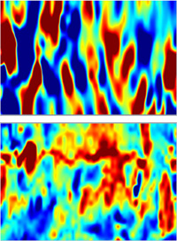 whose cover image is taken from the study. The co-senior authors were Louis Hodgson, Ph.D., associate professor of anatomy & structural biology, and Dianne Cox, Ph.D., professor of anatomy & structural biology and of developmental & molecular biology. Both are members of the Gruss Lipper Biophotonics Center.
As seen in the upper image, computational analysis "maps" the motion (y axis) of a 50-micron cell-edge section of a mouse macrophage over 10 minutes (x axis). Cyclic edge motion is visible in which protrusions (red areas) alternate with retractions (blue areas). The lower image of the same macrophage shows that protrusive activity correlates with Rac2 activity using fluorescent biosensors that reveal when and where Rac2 is activated in the cell (red = high Rac2 activity, blue = low).
Additional authors were Bin Wu, Ph.D., and Ph.D. students Veronika Miskolci and Yasmin Moshfegh, all at Einstein.
Other Top Stories
9/11 World Trade Center Exposure Linked to Heart Disease Among NYC Firefighters
On Becoming a Physician: New Einstein Students Receive White Coats and Stethoscopes
Novel Therapy for Acute Migraine Shows Promise in Phase 3 Clinical Trial
First Complete Wiring Diagram of an Animal's Nervous System
Multimillion Dollar NIH Grant to Help Reduce Opioid Use & Get Care to People Who Need It
NIH Grant Funds $23 Million Study of Diseases Affecting People Living with HIV
New TAILORx Data Guides Adjuvant Therapy in Younger Breast Cancer Patients
Einstein Celebrates Its 61st Commencement
Bolstering Biopsies: Testing Patients' Individual Cells to Guide Treatment
Tablet Blog