
Viewing Live Molecules
Dr. Robert Singer: Pioneering Biomedical Imaging for Viewing Molecules in Motion
For more than three decades, Dr. Robert Singer, professor and co-chair of anatomy and structural biology, co-director of the Gruss Lipper Biophotonics Center and professor of neuroscience and of cell biology, has been plumbing the mysteries of cells. For him and the researchers in the Singer lab at Einstein, this is the most basic and fundamental of life's questions. "The ultimate cell biology and consequently pathology," said Dr. Singer. "Is when you can describe which genes a particular cell is expressing at a particular place and time. If you know how the cell controls its genome in order to change its phenotype or behavior, you have the power to exercise control over that event."
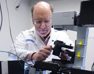
Robert H. Singer, Ph.D.Dr. Singer's persistent quest to look deeper and deeper into the lives of cells is paying off big, resulting in new discoveries and technologies that are giving scientists a previously unimagined ability to look inside living cells and watch them as the code of life is being transcribed.
The new technology, a powerful refinement of fluorescent in-situ hybridization (FISH), developed in Dr. Singer's laboratory almost 30 years ago, for the first time allows scientists to observe single molecules in single cells in real time – and is providing new understandings of how cells both live, and die.
"One of our major discoveries this year, by a graduate student, Tatjana Trcek, was that messenger RNA (mRNA) can be born with a built in ‘self-destruct' switch," said Dr. Singer. "That was one of the big questions – how do cells manage to degrade specific molecules at the right time, when they need to go away and let others take over? We found that the mRNA, as it is being made, assembles molecules on it that presage its own self destruction at a pre-determined moment during the cell cycle."
This once inconceivable ability to look into the molecules of living cells is the result of years of effort that have synergized resources, research, collaboration and technology to put Dr. Singer and his colleagues on the leading edge of innovation in a burgeoning field of medical research.
As a recent Science article, "How Cells Position Their Proteins," noted, pioneering research by Dr. Singer and others in the field on how mRNA is directed to specific destinations in cells was largely disregarded, or their results were perceived to be rare oddities. But, continuing research with the FISH technology developed by Dr. Singer has provided an overwhelming body of evidence that can no longer be ignored and is changing how scientists understand cellular function.
Dr. Singer's unique view of how to approach problems in cell research has benefited from excellent people, fruitful collaborations, a synergy of disciplines and technological leaps in microscopy. "Once we were able to tag RNAs with fluorescent molecules, we had to devise very sensitive detection systems, design optics so that we could image rapidly and develop ways to analyze the data – extracting the weak signals from the background haze," said Dr. Singer. "We were helped tremendously by the commitment of Einstein to build facilities to make this happen. Dean Spiegel, with generous support from Evelyn Lipper, made sure that an enormous amount of infrastructure and resources were put in place for this to succeed and to keep us at the forefront."
And the growth has been exponential. Shailesh Shenoy, director of engineering and operations at the Gruss Lipper Biophotonics Center and in the Singer lab, first came to Dr. Singer's lab in 1993 as a summer undergraduate student majoring in electrical engineering and biochemistry. "Improvements in reagents, optics, electronics and computation each contributed to the exquisite sensitivity of our microscopes that enable us to observe events, such as individual molecules of mRNA transiting the nuclear envelope, which would not have been possible even three years ago," said Mr. Shenoy.
He continued, "Einstein constructed several environmentally controlled rooms to permit these microscopes to operate with the high precision necessary to make quantitative measurements from which we track the position of an mRNA to within 30 nanometers every 20 milliseconds. In an hour or less, we now routinely perform computational analysis on microscope images that, in 1993, took 36 to 48 hours using less sophisticated algorithms."
With the accelerating pace of discovery, proliferating publications, and new funding, this has been a banner year for the Singer lab. And, with the technology, personnel and systems in place, the future is a boundless horizon. "There is no dearth of ideas," said Dr. Singer. "There are all sorts of questions we want to follow up. They require constant technology development."
Dr. Singer's latest interest lies in exploring mRNA in neurons. "Neurons form synapses at a point where they make contact, it is a discrete regulatory moment, and the mRNA must be available at this random moment in time. How is it regulated? The mRNA has to sit and wait a long, long time and then suddenly spring into action."
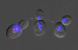
Messenger RNA molecules (in red) during cell divisionAnd, now that they have been able to put a single molecule detection system into place in living mice, Dr. Singer imagines a future where he can look in the brain of a living mouse and see what the genes are doing when the mouse is learning. "There is always a new angle, a new idea as soon as the technology becomes available" Dr. Singer said.
The ideas born in the Singer lab are being spread far and wide through ongoing and evolving collaborations and through the advancing careers of researchers and scientists who trained at the facility. "My experience in the Singer lab was extremely positive," said Dr. Yaron Shav-Tal, who was a postdoc in the Singer lab and is now a tenured faculty member at the Mina & Everard Goodman Faculty of Life Sciences & Nano-medicine Research Center at the Bar-Ilan Institute of Nanotechnology and Advanced Materials in Israel.
"Rob offers his postdocs a very high degree of scientific independence complemented by a nurturing environment in which to perform science using state-of-the-art technologies. This working atmosphere, in conjunction with friendly mentoring by Rob, drives students and postdocs to focus on fundamental scientific questions and, with hard work, results in important contributions to science."
Dr. Xavier Darzacq, also a former Singer postdoc who is now a faculty member heading the laboratory in Functional Imaging of Transcription at the Institut de Biologie de l'Ecole Normal, agreed. "The Singer lab provided decades of accumulated expertise and know-how. But, more than that, Rob's passion for science and his direct approach to problems is the most valuable aspect that helped my career."
Mr. Shenoy credits Dr. Singer with creating the ideal environment for experimentation. "Rob has created and maintained in his lab an environment in which people have fun doing science, discussing and testing hypotheses, developing technology and taking risks with their research goals," he said. "Rob enjoys doing science and surrounds himself with people who share his curiosity and passion for the research endeavor."
"I really am directed by curiosity," said Dr. Singer. "And, I'm intrigued by the randomness of biological systems. In basic research the premise is that knowledge is power. The more you know about how cells operate, the more you can do with them. The machinery that drives a cell has implications for all things cells do; how it is expressed in disease, how it is expressed in development. There are implications for things we can only begin to imagine."
View "Science Talk: Tracking mRNA in Living Cells" to hear from Dr. Singer about his research.
Posted on: Friday, February 24, 2012
Tablet Blog