
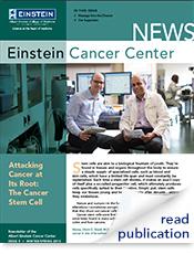
FULL STORY
Attacking Cancer at Its Root: The Cancer Stem Cell
Q: Are stem cells ever good for patients with cancer?
A: Yes. In fact, in some current therapies, “good” hematopoietic stem cells are first harvested from a donor’s blood or bone marrow. Then the patient is treated with intensive chemotherapy, and sometimes radiation therapy as well, to eradicate the cancer. However, this results in severe collateral damage to the patient’s normal bone marrow cells. To counteract this, the harvested stem cells
Stem cells are akin to a biological fountain of youth. They’re found in tissues and organs throughout the body to ensure a steady supply of specialized cells, such as blood and skin cells, which have a limited life span and must constantly be replenished. Each time a stem cell divides, it makes an exact copy of itself plus a so-called progenitor cell, which ultimately produces cells specifically suited to their location. Simply put, stem cells keep our tissues young and healthy decade after decade…unless they misbehave.
This article originally appeared in the winter/spring 2015 issue of the Albert Einstein Cancer Center Newsletter.Nature and nurture—in the form of genetic mutations and epigenetic alterations—sometimes corrupt stem cells, modifying their basic programs so that they churn out cancer cells instead of normal tissue cells.
Cancer stem cells were first observed in leukemias in the 1990s. They’ve since been found in many solid tumors, including breast, bladder, prostate, colon and liver cancers.
“The discovery of cancer stem cells and exploration of their self-renewal mechanisms have compelled us to take a fresh look at the basic biology of cancer and its treatment,” says Dr. Paul S. Frenette, director of Einstein’s Ruth L. and David S. Gottesman Institute for Stem Cell and Regenerative Medicine Research and a member of the Albert Einstein Cancer Center (AECC).
Cancer Therapy Revisited
For decades, oncologists have used two core therapies beyond surgery—chemotherapy and radiation—to shrink tumors, sometimes even putting cancer into remission. But more often than not, a few cancer cells survive and the disease returns. Some of those survivors appear to be cancer stem cells.
“Chemotherapy damages the DNA of rapidly dividing cells,” explains Dr. Ulrich G. Steidl, associate professor of cell biology and of medicine (oncology) and the Diane and Arthur B. Belfer Faculty Scholar in Cancer Research. “But cancer stem cells don’t divide very often, and therefore they resist these therapies. So in treating cancer, we’ve largely been trimming the branches of the disease while leaving the roots intact.”
Cancer Stem Cells and Acute Myeloid Leukemia
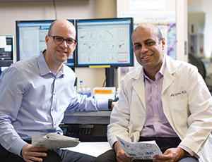
Drs. Ulrich G. Steidl (left) and Amit K. VermaDr. Steidl, leader of the AECC’s Stem Cells, Differentiation and Cancer Program, is trying to understand the roots of acute myeloid leukemia (AML), a cancer of the blood and bone marrow. Largely a disease of the elderly, AML takes about 10,000 lives a year in the United States.
Chemotherapy for AML is fairly effective against leukemia cells in the general circulation but has little impact on leukemia stem cells (LSCs), which reside in the bone marrow along with normal blood stem cells. Treatment also doesn’t touch preleukemic stem cells (pre-LSCs)—stem cells that have acquired some but not all of the alterations that make them cancerous.
“Pre-LSCs provide a silent reservoir for the return of LSCs,” says Dr. Steidl. “If we are to achieve a lasting cure for AML, we need to eliminate both LSCs and pre-LSCs. So it’s critical that we understand the earliest events in the transformation of a stem cell into a cancer stem cell.”
"In treating cancer, we’ve been trimming the branches while leaving the roots intact."
– Ulrich G. Steidl, M.D., Ph.D.
Molecule by molecule, gene by gene, Dr. Steidl is deciphering what makes good stem cells go bad. One culprit, it seems, is a gene called HLX. In a 2012 study published in Cancer Cell, he and his fellow researchers compared normal and precancerous bone marrow stem cells in mice. They found that HLX is expressed in abnormally high levels in pre-LSCs and LSCs. Elevated HLX expression was also found in bone marrow stem cells taken from patients with AML.
To prove that HLX is indeed a trigger for AML, the researchers showed that when they over-expressed HLX in healthy bone marrow cells, cancer-like blood cells developed. Conversely, when they inhibited HLX expression in leukemia cells in mice, the animals survived longer.
Dr. Steidl’s team is now trying to develop therapies that can block HLX activity in patients with AML. One approach is to disrupt HLX’s activity as a transcription factor: While one part of the HLX protein mediates its binding to DNA, another activates certain genes. “If we flood stem cells with dummy transcription factors—engineered peptides that contain the DNA binding but not the gene activation domain—we can crowd out the real ones and prevent oncogene expression,” he says.
For assistance in developing such peptides, Dr. Steidl has teamed with Dr. Evripidis Gavathiotis, assistant professor of biochemistry and of medicine (cardiology) and an expert in AECC’s Experimental Therapeutics Program. “Based on what was known about the transcription factor’s structure, we were able to synthesize a small peptide that can bind to DNA where HLX would normally bind, but without activating genes,” says Dr. Gavathiotis, who has also made the peptide more stable.
“Most synthetic peptides do not maintain their bioactive shape in aqueous environments such as the blood,” he explains. “Once that problem was solved, we observed that the peptide can penetrate LSCs and successfully slow their proliferation, at least in cell culture. We hope to move on to animal studies soon.”
Dr. Steidl has also found another gene implicated in AML. That gene, IL1RAP, is dysregulated early in human pre-LSCs and LSCs, and it controls a cell surface receptor. “When we knocked this gene down, we saw a near-complete absence of LSCs in both cell cultures and mice,” says the researcher, whose findings were published in Blood. Since the IL1RAP receptor sits on the cell surface, it promises to be a highly vulnerable target, says Dr. Steidl. He is now working with Dr. Gavathiotis to develop and test peptides that can interfere with the receptor’s function.
Cancer Stem Cells and Myelodysplastic Syndrome
"We found that genetically abnormal stem cells persisted in the bone marrow even when the disease was in remission."
– Amit K. Verma, M.B.B.S.
A second group of Einstein scientists, led by Dr. Amit K. Verma, professor of medicine (oncology) and of developmental and molecular biology, is studying the roots of myelodysplastic syndrome (MDS). MDS is a group of incurable blood disorders in which the bone marrow produces low numbers of red cells, white cells and platelets. Like AML, MDS is predominantly a disease of the elderly. About 15,000 new cases are diagnosed each year in the United States (about one-third of those patients will go on to develop AML). Treatment for MDS is limited to preventing or reducing complications, notably severe anemia.
The first indication that MDS is a stem cell disease came in 2012, when Dr. Verma, along with Dr. Steidl, found a variety of genetic and epigenetic alterations in the bone marrow stem cells of MDS patients, but not in stem cells of healthy controls. The researchers went on to show that these altered stem cells don’t function normally and can develop into abnormal blood cells.

Drs. Paul S. Frenette (left) and Evripidis GavathiotisIn the same study, the team followed the stem cells of an MDS patient during the whole course of his illness, from treatment to remission to relapse. “We found that genetically abnormal stem cells persisted in his bone marrow even when his disease went into remission,” says Dr. Verma. “And then, two months before he relapsed, the pool of abnormal cells expanded.” Other laboratories confirmed the findings, offering convincing evidence that stem cells are indeed the driving force behind MDS.
Researchers at Einstein and elsewhere found that a gene called STAT3, which has been linked to several types of cancer, appears to play a key role in MDS. The STAT3 gene codes for the STAT3 protein, which is necessary for stem cell survival. A clinical trial of pyrimethamine, a treatment originally developed for malaria that also inhibits the STAT3 protein, will begin soon at Montefiore under the leadership of Dr. Verma, chief of the division of hemato-oncology at Montefiore and co-leader of the AECC’s Stem Cells, Differentiation and Cancer Program.
"Cancer is complex and varied," says Dr. Frenette, also professor of medicine (hematology) and of cell biology at Einstein. "We hope that research on healthy and malignant stem cells will yield new approaches to target the cancer cells while preserving the healthy tissues."
Posted on: Tuesday, July 28, 2015
Tablet Blog